
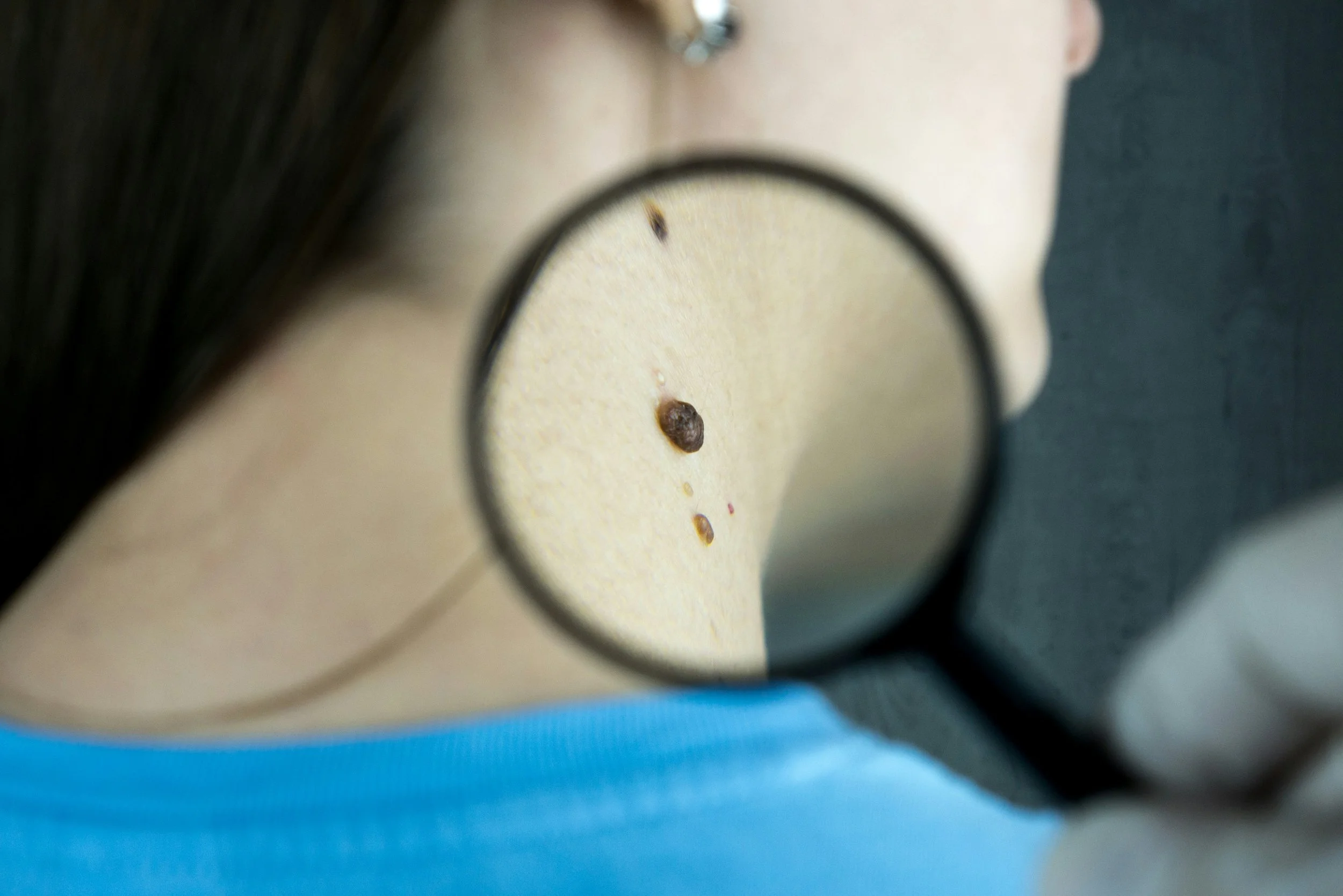
Skin lesions
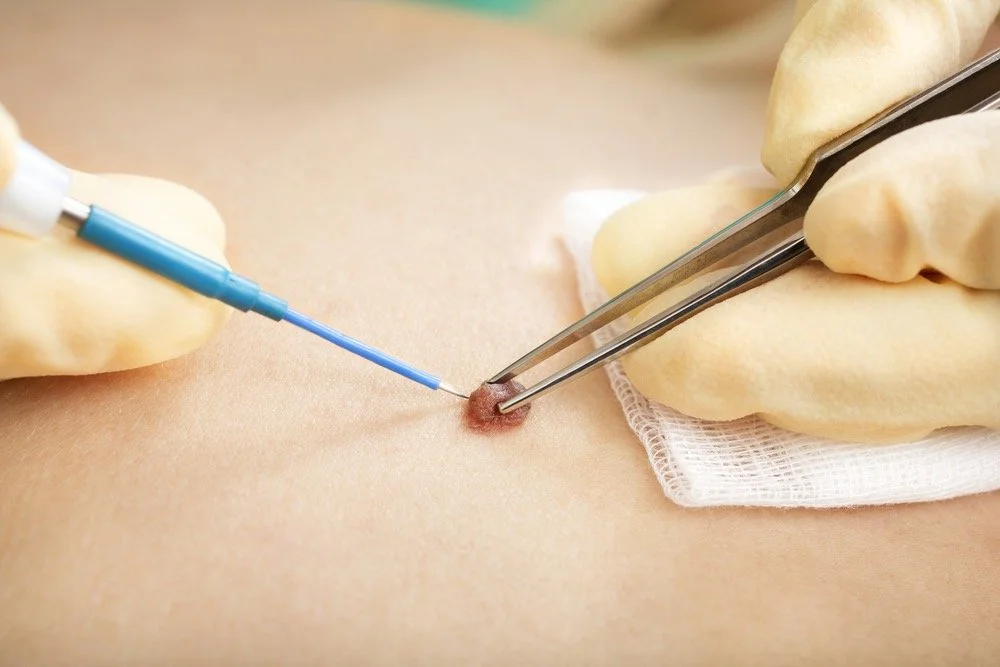
Skin lesion removal is a procedure to treat lumps or bumps on the skin such as moles, cysts, warts, or skin tags. These lesions are most often benign (non-cancerous), but may be removed if they are:
Painful or irritating
Cosmetically bothersome
Restricting movement
Showing changes in colour, shape or size, which may indicate a risk of skin cancer
Sometimes a lesion is removed to confirm a diagnosis. In these cases, the tissue is sent to the laboratory for analysis – this is called a biopsy.
-
Most procedures are carried out in the on-site operating theatre under local anaesthetic
You’ll usually be able to go home the same day
Removal methods vary depending on the type and location of the lesion. Mr Eccles may:
Shave the lesion level with the surrounding skin
Snip off small skin tags or soft lesions
Excise (cut out) the lesion completely, possibly with a margin of normal skin, and close the wound with stitches
In some cases, particularly for larger lesions or those in certain cosmetic or functional areas, reconstructive techniques may be needed after removal. These may include:
Local skin flaps – where nearby skin is rearranged to cover the wound
Skin grafts – where skin is taken from another part of the body and used to cover the area
Mr Eccles will advise if reconstruction is likely to be needed and explain the technique he recommends.
-
o You may feel mild discomfort as the anaesthetic wears off. Paracetamol is usually sufficient
Avoid aspirin or ibuprofen unless advised, as these may increase bruising
Keep the wound clean and dry – most dressings can be left in place for a few days
Avoid soaking the area in water (e.g. baths or swimming) until fully healed
If you’ve had a lesion removed from your face, sleeping slightly propped up can help reduce swelling
You’ll be advised when to return for stitch removal if non-dissolvable stitches were used
Avoid make-up on the wound until it is fully healed
Most wounds heal within 1–3 weeks, depending on the type of procedure
-
While generally a safe and straightforward procedure, all surgery carries some potential risks:
Scarring – most are small and fade over time, but some people are prone to hypertrophic or keloid scars
Bleeding, bruising, swelling or minor infection
Temporary or permanent numbness in the area, particularly in regions with many small nerve endings
Recurrence of the lesion – rare, but possible in some benign or cancerous lesions
Rarely, lesions thought to be benign may show signs of malignancy under the microscope. In these cases, further treatment or monitoring may be necessary
When reconstruction is required, there may be additional scarring, longer recovery time, or a need for further dressings and wound care
Mr Eccles will explain the specific risks and benefits in your case, and the steps taken to minimise complications.
-
Most removals are permanent
If your lesion is being removed due to cancer concern, a margin of healthy skin may also be removed and the sample will be analysed to ensure it is clear of any abnormal cells
You may be asked to return for a follow-up appointment if needed
Mohs surgery
Mohs micrographic surgery is a specialised technique used by dermatologists to precisely remove certain types of skin cancer, particularly on the face. It involves removing the cancer in thin layers while carefully checking each layer under a microscope until all abnormal cells are gone. This approach maximises cancer clearance while preserving as much healthy tissue as possible.
Mr Eccles works closely with expert dermatologists who perform Mohs surgery. Once the cancer is fully removed, he carries out the reconstructive surgery needed to repair the wound. This may involve fine suturing, skin grafts, or local skin flaps, depending on the size and location of the defect. The goal is always to achieve the best possible cosmetic and functional outcome.